 infobox@neuroproof.com
 +49 381 54345-660
ALS High Content Imaging Assays
NeuroProof Systems GmbH offers high-content screening assays with automated microscopy for the phenotypic screening of new ALS therapeutics. We deliver quantitative analyses of morphological biomarkers relevant to ALS. Based on its broad MEA experience with human iPSC-derived cell lines, NeuroProof Systems GmbH develops optimal imaging assays.
TDP-43 Mislocalization
The mislocalization of the TAR DNA-binding protein 43 (TDP-43) is a hallmark and early event in ALS. TDP-43 mislocalization from the soma toward the cytoplasm is part of the TDP-43 proteinopathy. TDP-43 is a strong phenotypic biomarker in ALS pathology, connected to a broad spectrum of pathomechanisms.
We have detected in C9orf72 cells mislocalized proteins of TDP-43:
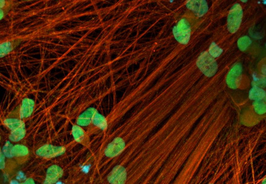
Image: Control spinal motor neurons with TDP-43 in somas only. TDP-43 green, β-III tubulin red, DAPI blue.
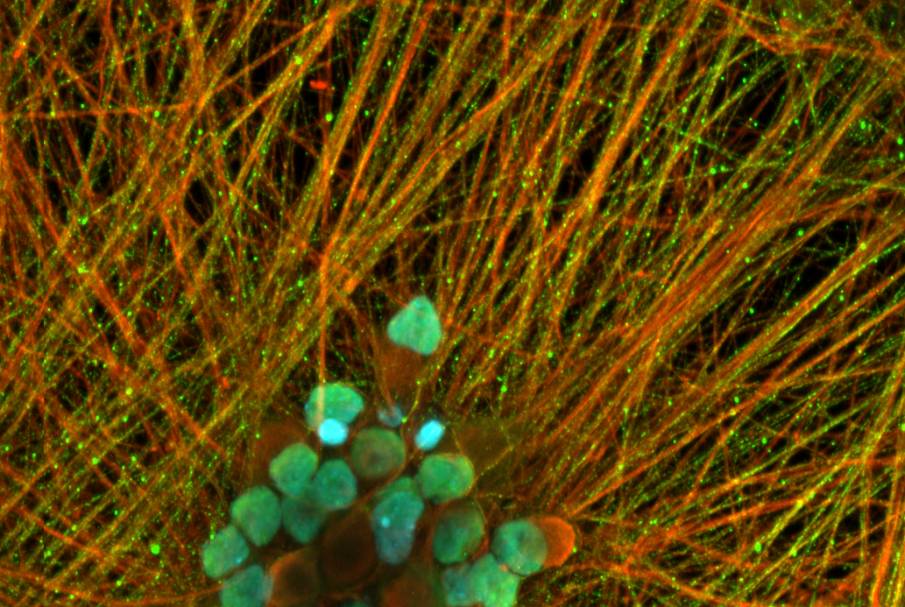
Image: C9orf72 spinal motor neuron cells with TDP-43 in somas and axons. TDP-43 green, β-III tubulin red, DAPI blue.
TDP-43 mislocalizations can be reliable and highly reproducible, detected, and quantified. Inhibitors of TDP-43 demonstrate a nice dose-response dependency of TDP-43 clearance. This assay can be outperformed in 96 well plates and is suited for cost-effective and comprehensive projects.
In our hands, the adenosine 2A agonist, JMF-1907, and the sigma receptor agonist, PRE-084, demonstrated an apparent reduction of TDP-43 mislocalization in C9orf72 cells..
Dipeptides
The dipeptide repeat protein Poly(GR) in C90rf72 cells is responsible for TDP-43 pathology, and it impairs protein translation and forming of stress granules. Therapies targeting the lowering of Poly(GR) are a potential approach for new ALS therapies.

Image: Control spinal motor neuron cells with Poly(GR) staining. Poly(GR) green, β-III tubulin red, DAPI blue.

Image: Control spinal motor neuron cells with Poly(GR) staining. Poly(GR) green, β-III tubulin red, DAPI blue. Poly(GR) particles are forming speckled dots in the soma.
Stress Granules
Stress granules (SG) are non-membrane-bound RNA-protein granules that assemble through phase separation in response to cellular stress. SGs are composed of RNA-binding proteins RBPs. Persistent structures of stress granules are seeding germs of RNA-binding proteins, which leads to protein aggregation and mitochondrial damage in ALS.
G3BP1
G3BP1 is an essential scaffold protein of stress granules, which we use as a marker for SGs.

Image: G3BP1 staining of spinal motor neurons, G3BP1 green, fixed at div 15.
Automated Imaging with 96 well plates
We perform our imaging projects with an automated imaging system in 96 well plates or as a supplementary study on transparent MEA plates.
Intelligent Quantitative Image Analysis
AI-driven intelligent image analysis delivers robust and reliable results. For example, we perform projects with immunostaining fluorescence microscopy or light microscopy to assess neurite outgrowth.
Get in Touch
NeuroProof is continuously expanding its assay portfolio, asking us for your ALS high-content imaging campaign. High-content Imaging is an effective entry point to your ALS projects..